Introduction
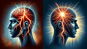
Migraine headaches are a neurological disorder that affects approximately 12% of the global population, with women being three times more likely to experience migraines than men. Characterized by severe head pain, nausea, vomiting, and sensitivity to light and sound, migraine attacks can be incredibly debilitating, resulting in lost productivity and diminished quality of life. While migraines have been documented for centuries, the underlying pathological mechanisms behind migraine attacks have remained elusive. However, new research developments are starting to shed light on the complex neurological roots of migraines, paving the way for more targeted and effective treatments.
A migraine attack is understood to originate from abnormal neurological activity causing dilation of blood vessels and the release of inflammatory substances that irritate pain-sensitive nerve fibers. This leads to activation of the trigeminal nerve, the primary pathway for pain signals from the face and head. The trigeminal nerve transmits signals to the brainstem, which relays them to higher cortical regions involved in processing pain perception. One prominent theory is that migraines are triggered by cortical spreading depression (CSD), a slow-moving wave of neuronal hyperactivity followed by suppressed brain activity thought to correspond with the visual aura some patients experience. While CSD and activation of the trigeminal nerve pathway are key elements of migraine pathophysiology, emerging research indicates that migraines involve multiple brain regions and regulatory systems.
New insights into the complexity of the migraine brain offer hope for better management of this debilitating disorder. This article will review recent discoveries on the role of the hypothalamus, brainstem, thalamus, and higher cortical regions in migraine attacks and symptomology. The implications of these neurological insights for future migraine treatment and prevention will also be discussed. Elucidating the underlying neurological aberrations in migraine is key to developing new targeted drugs and interventions to more effectively treat and potentially prevent migraine episodes.
New insights into the role of the hypothalamus in migraine
The hypothalamus is a small brain region that plays key roles in regulating pain, sleep, appetite, hormones, mood, and other critical bodily functions. Historically, migraine research has focused on the brainstem and cortical regions, but new evidence indicates the hypothalamus may actually play an orchestrating role in migraine attacks.
Recent studies utilizing advanced brain imaging techniques have found that the hypothalamus exhibits significantly increased activity during the premonitory phase in the 24 hours before a migraine attack. The hypothalamus remains hyperactive throughout the duration of the attack as well. This hypothalamic hyperactivity correlates with activation of the trigeminal nerves and development of pain.
In particular, the posterior region of the hypothalamus shows the greatest activation, especially in the premonitory stage. The posterior hypothalamus contains networks of neurons that regulate functions like sleep, mood, and craniofacial pain that are often disrupted in migraine patients. Hyperactivity in this region could help explain symptoms like sleep disturbances, food cravings, nausea, and head pain that precede an attack.
These findings indicate the hypothalamus is potentially the central hub that initiates migraine episodes and maintains the cascade of neurological and vascular changes that generate head pain and other symptoms. The hypothalamus may trigger CSD and activation of the trigeminal pathway. Treatments targeting hypothalamic hyperactivity and dysregulation could potentially prevent or stop migraine attacks at their source.
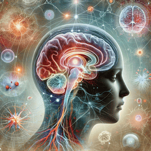
Other brain regions involved in migraine
While the hypothalamus appears to play a key orchestrating role, migraine is a complex neurological disorder that involves multiple brain regions. Research has also highlighted the contributions of the brainstem, thalamus, and higher cortical areas to migraine pain processing and symptoms.
The brainstem receives signals from the trigeminal nerve and contains networks of neurons that modulate pain perception. Brain imaging studies show that migraine patients exhibit sustained hyperactivity in certain brainstem nuclei during and between attacks. This brainstem hyperactivity positively correlates with the intensity of head pain.
The thalamus serves as a relay hub connecting sensory pathways to the cortex. Neuroimaging evidence indicates the thalamus shows abnormal activation levels during migraine episodes, potentially disrupting normal sensory processing.
Cortical regions like the somatosensory cortex, insula, and cingulate cortex are involved in pain perception, interoception, and modulation of pain signals. Some studies report that migraine patients exhibit atypical activity in these areas in response to experimental pain compared to healthy controls. Abnormal cortical excitability and processing likely contributes to migraine pain and other symptoms like light and sound sensitivity.
The amygdala and hippocampus also show altered activity patterns during migraine attacks, which could underlie associated mood disturbances and cognitive complaints like brain fog. Overall, the emerging picture is that dysfunction across interconnected brain networks involved in sensory processing, pain modulation, and homeostatic regulation contributes to the mosaic of migraine symptoms patients endure.
Implications of new research for migraine treatment and prevention
These neurological insights set the stage for new targeted treatments that directly address the underlying brain disturbances driving migraine episodes.
One promising area is therapies that inhibit hypothalamic hyperactivity and regulate dysfunctional networks in the brainstem and thalamus. Newly developed neuromodulation techniques like transcranial magnetic stimulation (TMS) and neurofeedback allow non-invasive and focused modulation of activity in migraine-associated brain regions. Early trials of TMS targeting the occipital cortex shows potential for both treating acute migraine attacks and reducing recurrence when applied preventatively.
Understanding the neurological underpinnings of migraine also helps identify new drug targets. A class of drugs called CGRP monoclonal antibodies act to block the release of calcitonin gene-related peptide, a molecule involved in trigeminal nerve activation and pain signaling. In clinical trials, CGRP inhibitors significantly reduced migraine frequency in chronic sufferers. Similar biologic drugs targeting dysfunction in brain pathways upstream of the trigeminal nerve may yield even better results.
While more research is needed, uncovering the neurological roots of migraine brings hope for transforming migraine treatment. Meticulous characterization of the migraine brain will enable physicians to apply personalized, mechanism-based therapies to better prevent and manage migraine for their patients. Catching and treating migraine attacks early is also key to preventing progression to chronic and debilitating migraine. Integrating the latest neurological insights stands to significantly improve quality of life for the millions of migraine sufferers worldwide.
Conclusion
Migraine is an enormously prevalent and disabling neurological disease involving dysfunctional brain networks that regulate pain, sensory processing, and homeostasis. While prior migraine research focused heavily on the trigeminal nerve and cortical spreading depression, emerging evidence elucidates the involvement of multiple interconnected brain regions, including key roles of the hypothalamus, brainstem, and thalamus. New insights into the neurological underpinnings of migraine will enable more targeted treatments aimed at rectifying the brain disturbances that generate migraine episodes. Continued research bringing neurologists closer to a complete map of the migraine brain offers new hope for transforming migraine care and reducing the global burden of this debilitating neurological disorder.
References:
- Schulte, L. H., Allers, A., & May, A. (2017). Hypothalamus as a mediator of chronic migraine. Neurology, 88(20), 2011-2016.
- Noseda, R., & Burstein, R. (2013). Migraine pathophysiology: anatomy of the trigeminovascular pathway and associated neurological symptoms, CSD, sensitization and modulation of pain. Pain, 154, S44-S53.
- Stankewitz, A., Aderjan, D., Eippert, F., & May, A. (2011). Trigeminal nociceptive transmission in migraineurs predicts migraine attacks. The journal of neuroscience, 31(6), 1937-1943.
- Moulton, E. A., Becerra, L., Johnson, A., Burstein, R., & Borsook, D. (2014). Altered hypothalamic functional connectivity with autonomic circuits and the locus coeruleus in migraine. PLoS One, 9(4), e95508.
- Lippi, S., Mattioni, A., & Toldi, J. (2019). Neuropeptides in the hypothalamic supraoptic and paraventricular nuclei in migraine animal models: a mini review. Frontiers in cellular neuroscience, 13, 214.
- Noseda, R., & Burstein, R. (2013). Migraine pathophysiology: anatomy of the trigeminovascular pathway and associated neurological symptoms, CSD, sensitization and modulation of pain. Pain, 154, S44-S53.
- Stankewitz, A., & May, A. (2011). Increased limbic and brainstem activity during migraine attacks following olfactory stimulation. Neurology, 77(5), 476-482.
- Schwedt, T. J. (2014). Chronic migraine. BMJ, 348.